In the last blog we touched on some pertinent diagnostic features that we are able to see from a two dimensional panoramic x ray. We will continue this discussion in this blog.
A panorex enables one to:
1) Identify fractures of the teeth and jaw that may occur as a result of trauma, i.e. a car accident. This type of x ray is especially helpful if an individual is unable to open their mouth, as the machine circles around their head… Also valuable for patients who gag and cannot tolerate an intra oral x ray.
2) Ensure adequate ‘setting’ of facial bones (by a surgeon) that may occur as a result of any trauma, as mentioned in number one.
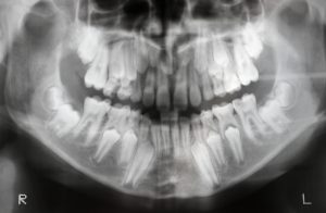
3) Analyze eruption patterns of both the primary and adult teeth. A panorex enables us to see both sets of teeth in one image. Note: The above image is an example of this. In this panorex one can even see the early formation of the wisdom teeth. They look like little ‘buds’ at each of the four corners of the jaw.
Question: How old do you think this patient is ?
Answer: I would guesstimate that they are at least 10 years old. This is based on the fact that the individual’s first permanent molars are erupted but their second permanent molars aren’t. The first molars erupt at about 6 years of age and the second at 12 years old. And the first permanent molars in this individual look fully mature; it’s not as if they just erupted into the patient’s mouth…if this was the case, the individual would be about 6 years old.
4) Any signs of oral cancer. This may be the result of metastases of cancer from other areas i.e. the breast, lung, prostrate, thyroid gland, kidney and colon.
5) A malignancy, as mentioned in number four, may cause paresthesia or altered nerve sensation of the lip. This may be identified in a panorex as a malignancy compressing the major nerve that runs below the teeth in the lower jaw.
6) To a certain extent it may be helpful to diagnose Temporomandibular Joint Disease i.e. one may be able to see arthritic changes in the ‘joint’ itself. For diagnosis of any damage to the joint it is better to rely on an MRI or CAT scan which will provide a three dimensional view as opposed to the two dimensional one provided by a panorex.
7) Analysis of the Maxillary Sinuses. These are actual spaces on either side of the upper jaw just below the cheek bones. Many times the roots of the upper molar teeth project into the sinuses…when the sinuses become congested, this often translates into an individual feeling pressure in the associated teeth especially upon clenching. For more information regarding the maxillary sinuses (and how they can effect implant placement), please read the blog “Sinus Lift…Really?” posted on March 25th, 2012.
8) For the diagnosis of any genetic and/or developmental abnormalities such as Cleft Palate.
9) Detecting the presence of periodontal disease i.e. disorders of the gum and bone. This is possible with a panorex, however a Periapical x ray is more desirable for this purpose. For more information of the diagnostic value of a peri apical x ray please review the blog “Peri Apical X Rays,” posted on May 20th, 2013.
We’re almost finished our discussion of the panorex x ray, however there is one more very important diagnostic and lifesaving feature that a panorex may convey……please stay tuned!
Dr. F. Keshavarz Dentistry


